Yale Engineer Leverages Cutting-Edge Technology for Fibrosis Detection

Associate professor of biomedical engineering Rong Fan has begun a $1.85 million project intended to create a minimally invasive method for the detection and measurement of bone marrow fibrosis, a pathological state wherein the affected organ forms an excess amount of fibrous connective tissue, causing severe clinical symptoms such as systemic inflammation, elevated white cell counts, and enlarged spleen. The method will build on single-cell analysis technology previously developed by Fan.

As part of a consortium of researchers funded by the National Institute of Diabetes and Digestive and Kidney Diseases (NIDDK), the project will contribute to discovering a common mechanism for the development of fibrotic pathology in the bone marrow, kidney, liver, prostate, and urinary tract. Fan’s research, the only project in the consortium to study fibrosis in bone marrow, will closely examine individual cell secretion of cytokines, a broad category of small proteins used for cell signaling — including signals that cause inflammation and wound repair.
“Identifying what cytokines are being produced to mediate the healing process tells you if either the cells are normal and will heal the wound or if the cells have acquired abnormal functions, probably due to mutation, and will generate fibrotic scar tissue,” said Fan. “But as we move beyond just distinguishing ‘normal’ from ‘mutated,’ we hope to discover what cytokines develop as the pathogenesis changes from early chronic inflammation to fibrosis, a knowledge that not only elucidates the cellular mechanism but could also lead to early detection of the pathological changes toward fibrosis.” He also added that the project was additionally benefited by Yale’s history of excellence in non-malignant hematopoiesis research, including work done by Diane Krause of Yale Stem Cell Center.
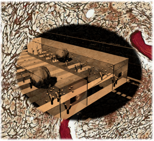
Foundational to the grant, therefore, is Fan’s nanoscale proteomic technology, a microchip platform for highly multiplexed protein measurement in single cells that can be potentially combined with a microfluidic processor to facilitate genomic, epigenetic, and transcriptional profiling at the single-cell level. Using only very small quantities of cells, Fan’s device is capable of highly multiplex detection of more than 40 cytokine secretions at the single-cell level to distinguish minute functional differences in otherwise similar cells.
Fan’s device will be used to analyze human bone marrow from patients identified by co-principal investigator Dr. Ross Levine of the Memorial Sloan-Kettering Cancer Center. The team will perform single-cell cytokine profiling on bone marrow cells including specific subpopulations in order to identify the unique cell types that show the most contrast between normal and abnormal cells with potential to initiate fibrosis. While this project is largely focused on the measurement of bone marrow cells, the technology has the potential to be further developed into a method for using the more-commonly available blood samples to detect and profile abnormal cytokine-secreting cells shed from bone marrow into the blood.
“With this device, our first goal is to identify new cellular targets for therapeutic intervention in bone marrow fibrosis,” said Fan. “But because these cytokine functions are a common mechanism in a range of human pathologies, the research is an important step toward creating a generic platform to diagnose other organ fibrosis diseases.”
The NIDDK, the fifth largest Institute at the National Institutes of Health, conducts, supports, and coordinates research on the diseases of internal medicine and related subspecialty fields, as well as many basic science disciplines. Other projects funded by the NIDDK Fibrosis Consortium are from Massachusetts General Hospital, Dana Farber Cancer Institute, and the Mayo Clinic.

