
Students in Medical Devices Class 'Innovate With Purpose'
Few presentations are likely to demonstrate a new laparoscopic surgical technique, let alone follow that demonstration with a game that allows nurses and doctors to compete for who can perform the best CPR. But for this year’s Medical Device Design & Innovation course, that was only half of the final presentations.

Team-taught by Joe Zinter of the School of Engineering & Applied Science and Jean Zheng of the Yale School of Medicine, the course brought together interdisciplinary teams of students to address unmet clinical needs identified by physicians from the Yale School of Medicine and Yale-New Haven Hospital.
“Fifteen weeks ago,” said Zinter, associate director of the Center for Engineering Innovation & Design, “five physicians representing four exciting challenges asked our class for better ways of repairing hernias and better ways of helping dysphagic patients swallow, better ways of teaching CPR and better ways of training healthcare professionals to use external pacemakers.” Those physicians, he added, then served as both clients and mentors, guiding the students as they developed and refined their solutions.
“The students knew that this was going to be a challenging course,” said Zheng, engineering director for the Center for Biomedical and Interventional Technology, “but they wanted this unique opportunity to make a difference in patients’ lives, and that’s what matters.”

Learning to Keep Pace
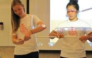
The first presentation of the night began with a heart attack. “Andrea,” we were told, was a ten-year-old girl played by senior biomedical engineering major Andrea Torres; she had just come out of a successful heart surgery, but was feeling weak and tired. Soon, Dr. Alex (played by junior electrical engineering major Alex Carrillo) had diagnosed her with bradycardia—an abnormally slow heart rate—and so attached an external pacemaker in an attempt to stabilize the patient. But it turned out Dr. Alex didn’t have any experience using the machine, and neither did his nursing staff. “Oh no!” he said. “What can we do?”
The team’s comedic dramatization showed that while external pacemakers are a common piece of hospital equipment, it can be difficult to get experience using the device—a difficulty caused, according to mentor Dr. Stephanie Sudikoff, by the absence of an appropriate platform for hands-on training with the device.

In response, the Sim Team created a small device named the CAAAM4000 that can simulate the electrical waveforms (both healthy and unhealthy) typically sent from the heart to the patient’s heart-rate monitor; as well, the waveforms can also be modified with input from the external pacemaker. In this way, the team’s device provides physicians the opportunity to practice using the external pacemaker to influence the patient’s cardiac patterns in real time—an invaluable experience that isn’t otherwise possible.
“Whereas you’d normally have a heart connected to the pacemaker and the monitor,” said Carrillo, “our heart is these four little knobs and an iPhone app.” In addition to having already tested the device with clinicians at Yale-New Haven Hospital, Torres, Carillo, junior electrical engineering major Christopher Datsikas, biomedical engineering graduate student Maxwell Emerson, and junior mechanical engineering major Amy Rockwood plan to continue developing a training program to accompany their invention.
“We have previously had to learn and practice on live patients,” said Sudikoff, associate clinical professor of pediatrics critical care and SYN:APSE Simulation Center medical director. “You have filled a vacuum.”

Better Than a Spoonful of Sugar
Of the four teams in the class, only the second team’s invention had the additional requirement that it appeal to, and be usable by, patients with perhaps no clinical experience—a necessity since the device would be used almost entirely outside of a hospital and entirely controlled by the patient without medical supervision.
The problem was swallowing: When you swallow food or drink, pressure sensors in the back of the throat activate the anterior digastric muscles to lift the larynx and lower the epiglottis, which prevents the food or drink from entering your windpipe. However, between ten and fifteen million people—especially geriatric and stroke patients—lose their ability to fully control the anterior digastric muscles, a condition known as dysphagia that makes swallowing difficult or impossible. Patients with dysphagia often develop depression or malnutrition, and the only effective cure—aside from physical therapy for very mild cases—is to begin using a feeding tube, a sometimes painful procedure that can increase social isolation and significantly lower the quality of life.

Team Uplift counteracted these negative effects by designing a wearable user-operated neck collar that uses a small electrical current to stimulate the muscles in the throat, thereby lowering the epiglottis. Built using an Arduino microcontroller, a rechargeable battery, and radio and Bluetooth receivers for activation and calibration, the entire mechanism fits on a 2 inch by 2 inch custom-built circuit board easily installed in a foam remote-controllable collar. For operation, the wearer need only push a button while swallowing and the electric current will contract the muscle to raise the larynx and lower the epiglottis.
The team, which includes senior biomedical engineering major Brianna Chrisman, junior history major Rebecca Delman, junior mechanical engineering major Benjamin Rosenbluth, junior economics major Paul Singer, and senior electrical engineering major Alejandro Villarreal, hopes to further reduce the size of the electronics and collar while refining the waveform parameters to be tunable to each individual patient’s muscular response.
“Dysphagia is a very complex process,” said Dr. Boris Paskhover, the team’s physician mentor and a resident at Yale School of Medicine, “and this team did a fantastic job targeting the small muscles that cause swallowing.”

Putting the Super in Superglue
The third team began their presentation with a literal—though rather quiet—bang, as second-year medical student John Gaudet used a tack gun to “repair a hernia” on a cantaloupe held by senior biomedical engineering major Thomas Morley. As Gaudet shot the spiral metal tacks into the fruit, he explained that hernias—which are the protrusion of an organ through the wall of the cavity that normally contains it—are usually fixed with a mesh that is sutured and tacked into place over the tear in the cavity wall.
However, the sutures and tacks that rip through muscle wall and peritoneum lining—as many as thirty tacks are employed in a repair—can cause severe patient discomfort that necessitate prolonged immobilization during recovery. Enter team mentor and associate professor of surgery Dr. Kurt Roberts, who proposed that the team find a less-painful method for performing the surgery.

To accomplish this task, Gaudet, Morley, junior mechanical engineering major Genevieve Fowler, senior biomedical engineering major Joyce Li, and junior biomedical engineering major Thomas Ryan designed a silicone-bordered mesh that without any sutures or tacks can be applied using only sterilized superglue. Once positioned in the body, a small tube embedded in the silicone opens on one end of the mesh while a tube on the other end vacuums the air, pulling the superglue across the surface and suctioning the mesh into contact with the perforation.
Additionally, the materials are ideal for minimally-invasive laparoscopic deployment: because the silicone is molded to be only 1mm thick, the entire tool can be rolled up and delivered through a laparoscopic port; once inside the body, the mesh can be unrolled by inflating the perimeter tubes with air.
The Hernia Team, however, did not just rely on words but in fact showed the efficacy of their innovation through a live laparoscopic surgery performed on a simulated “patient.” Using the same tools that would be used in an actual hernia repair, the team deployed their device and skillfully attached it to the inside of their simulated patient’s gut.
“This presentation isn’t just a proof of concept,” said Li, “it’s a successful surgery.”
Staying Alive, Staying Alive
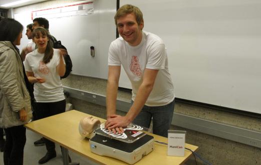
The night came to a rousing finale with Team ManiCan, who designed a next-generation manikin for improved cardiopulmonary resuscitation (CPR) training. CPR is performed by externally pumping the heart during cardiac arrest to maintain the flow of oxygenated blood through the body and manually preserve intact brain function; this procedure extends the brief window of opportunity for heart resuscitation using a defibrillator.
Co-mentored by associate professors of pediatrics emergency medicine Drs. Melissa Langhan and Marc Auerbach, Team ManiCan sought to help clinicians achieve greater consistency in their CPR performance, which requires a compression rate of 100-120 compressions per minute (roughly the speed of The Bee Gee’s “Staying Alive” or Carly Rae Jepsen’s “Call Me, Maybe”) at a consistent compression depth and recoil of 2 to 2.5 inches—a depth necessary to push out and refill the heart’s full blood capacity.
Though many CPR training manikins are already available, the team—consisting of senior biomedical engineering major Katelyn Chan, junior mechanical engineering major Jeffery Gau, junior cognitive science major Aaron Gertler, senior computer science major David Mandelbaum, and junior mechanical engineering major Patrick Wilczynski—created a manikin that offers unique real-time visual feedback through an exposed tube that displays the flowing “blood,” which is actually a mixture of glycerin and potassium iodide for proper consistency, and glitter for an eye-catching visual cue.

But to improve not only one individual’s CPR proficiency but an entire medical department’s, the team also incorporated sensors to model the relative concentration of CO2 in the bloodflow—a measure of direct physiological response to CPR that incorporates data from compression rate, depth, and recoil to deliver a single numerical score. This score is delivered in real time to a web-based software with a user interface that has been optimized for easy dashboard display of each data variable, an appearance that resembles a speedometer that has been color-coded to highlight the ideal range.
Equally crucial to the team’s intentions, however, is how the software records and displays the overall CPR data: the effectiveness of the CPR is measured over a period of time, with a single value posted at the end to a leader board. This easy comparability of scores enables a user to track improvement while also promoting healthy competition among clinicians attempting to get the highest score. “We envision a scenario,” said Mandelbaum, “where doctors and nurses would be in break room, practicing CPR and competing with each other as a motivational tool.”
After an invigorating thirty-second exhibition of how the manikin and software work, the teams all finished with a pause to catch their breath, allowing space for SEAS Deputy Dean Vincent Wilczynski to sum up the night. “These projects all displayed a remarkable level of innovation and technical expertise that is on par with any design class in the world,” he said. “However, the opportunity to develop these projects in collaboration with clinicians at the Yale School of Medicine is a fantastic opportunity that doesn’t just encourage innovation—it’s innovation with a purpose.”